Attic Cholesteatoma Radiopaedia

Cholesteatoma Radiology Reference Article Radiopaedia Org

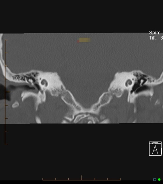
Fd Acquired Pars Flaccida Cholesteatoma Left Coronal T Bone Ct Image Shows An Atticoantral Nondependent Homogeneous Soft Radiology Image Shows Head And Neck

Mastoditis Middle Ear Head And Neck Sinusitis

Cholesteatoma Radiology Case Radiopaedia Org
Acquired Cholesteatoma Radiology Reference Article Radiopaedia Org

Cholesteatoma Radiology Case Radiopaedia Org
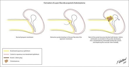
If untreated a cholesteatoma can eat into the three small bones located in the middle ear the malleus incus and stapes collectively called ossicles which can result in nerve deterioration deafness imbalance and vertigo.
Attic cholesteatoma radiopaedia.

Image Result For Scutum Erosion Facial Nerve Eustachian Tube Dysfunction Middle Ear

Mastoditis Middle Ear Head And Neck Sinusitis
Pars Tensa Cholesteatoma Radiology Case Radiopaedia Org

Ct Through The Orbits Obtained Initially Without Contrast And Then With Contrast While The Patient Performed A Valsalva Manoeuvre In The Kt Ppn
Source : pinterest.com
